
Screening methods for breast cancer
The fact that breast cancer is common, its frequency is increasing, that it can be treated at an early stage and that diagnosis at an early stage is possible in today’s conditions increases the importance of screening methods in breast cancer.

The screening method is decided according to the patient’s age, breast structure, and physical examination findings.
CLINICAL BREAST EXAMINATION
Breast examination is performed by a specialist doctor. In general, it should be done 1-3 years before the age of 40, and every year after the age of 40. The frequency of examination is determined by the doctor according to the presence of risk factors and the symptoms detected in the breast.
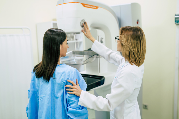
MAMMOGRAPHY
Mammography is the most important imaging method used in early diagnosis.
Mammography is a special breast X-ray film taken with low doses of X-rays. It is performed to detect the formations in the breast that are too small to be detected by examination, especially those that are indicative of cancer, and to see the breast details and internal structure of women with fibrocystic structure.
No medication is used during mammography. No injections are given. The breast is gently compressed between two plates. The purpose of compression is; The aim is to increase the image quality by preventing the movement of the breast, to ensure that a smaller amount of X-ray is received by reducing the thickness of the breast, and to prevent erroneous results by opening the formations that may overlap in the breast.
With the new generation digital mammography, the amount of X-ray exposure has been significantly reduced.
Mammography is performed horizontally and vertically.
There is no need for preliminary preparation for mammography. There is no change in the image taken depending on the menstrual period. However, in terms of sensitivity, it may be more comfortable to have it taken outside the menstrual period.
She is naked above the waist during the shooting. It is recommended not to apply deodorant, talcum powder or lotion to the breast and armpit area in order not to affect the appearance.
BIRADS (BREAST IMAGING REPORTING and DATA SYSTEMS) BREAST IMAGING AND REPORTING SYSTEM
It is a reporting technique that shows the path to be followed as a result of mammography.
BIRADS 0: It is a situation where something is seen on mammography but it is not clear what it is. Additional examinations are needed.
BIRADS 1: means the mammography result is normal.
BIRADS2: It is a situation where something is seen on mammography but it is 100% sure that it is benign.
BIRADS3: This is a condition thought to be very likely benign. Follow-up after 6 months is recommended.
BIRADS4: These are cases where the result must be supported by biopsy. This is a situation in which the mass seen is likely to be cancerous.
BREAST ULTRASONOGRAPHY
It is an effective method used in women with dense breast structure. It is effective in young women and women with familial risk, in cases where mammography is normal but there is a palpable mass. It is effective in detecting formations that cannot be seen in mammography when tissues overlap.
Ultrasound does not contain radiation. In cases of a palpable mass in women under the age of 40, ultrasonography is preferred first.
A small amount of pain may be felt during periods when the breast is sensitive. Ultrasound is preferred for signs of infection in the breast (pain, swelling, redness). /p>
In cases of suspicious findings during mammography or clinical breast examination, ultrasonography is preferred for additional insight.
MAGNETIC RESONANCE MRI
Magnetic resonance is the process of obtaining images using radio waves in a strong magnetic field created by large magnets. It does not contain radiation. It is used to detect formations and differences that do not exist in the natural structure of the body. It gives good results in imaging soft tissues.
MRI detects 20%-25% of foci that cannot be detected by mammography, ultrasonography and manual examination. Especially in dense breasts with fibrocystic structure, MRI plays a role in clarifying the surgical treatment plan. It is a guide in cases where it is thought that cancer may recur.
When cancer is detected in one breast, it is often used to look for formations that indicate cancer in the same breast or the other breast.
It is not generally used as the first step in screening. In some cases, MRI is used to support ultrasound. It is also requested in addition to mammography in high-risk patient groups. MRI is not affected by the density and structure of the breast.
It is also used for control purposes after breast cancer treatment and to measure the effectiveness of chemotherapy in advanced breast cancer.
BREAST BIOPSY
When a suspicious lesion is seen in the breast with imaging methods, scans at the cell structure level are required to understand whether it is cancer or another formation. Taking a sample suitable for evaluation with the help of various instruments and devices under the guidance of radiology is called breast biopsy.
The lesion may be seen better on mammography, ultrasonography or MRI. A biopsy is performed using whichever method provides a better image. Sometimes a biopsy can be performed surgically.
The sample taken is examined in the pathology laboratory. It is reported whether there is cancer or not. The purpose of biopsy is to take as many samples as the pathologist can evaluate.
There are different methods of biopsy. The radiologist decides on the appropriate technique based on the amount of sample to be taken, the breast structure and the condition of the lesion.
During the biopsy, clothing above the waist is removed. The area to be sampled is anesthetized with a technique called local anesthesia, which numbs only that area. It is aimed that you do not feel any pain during the procedure. You may have pain during the day or the next day after the procedure. You can use painkillers according to your doctor’s recommendation. After the procedure, the sampled area will be closed. This area needs to be monitored for bleeding.